MRI Imaging for Prostate Cancer
Magnetic resonance imaging (MRI) is an important tool in diagnosing and treating prostate cancer. It uses magnetism and radiowaves to create a picture of what’s inside your body. A positive biopsy is required to definitively diagnose prostate cancer.
MRI imaging aids the precise targeting of areas for focal therapy. Recent developments in MRI technology have allowed radiographers to create more accurate scans. This includes the development of multiparametric MRI scans (mpMRI).
What is a Multiparametric MRI (mpMRI) scan for prostate cancer?
A multiparametric MRI (mpMRI) is a detailed prostate scan that combines several imaging sequences to locate and grade cancer before any biopsy. Used first in the pathway, it reduces unnecessary biopsies and improves accuracy — MRI-targeted biopsy detects 36.3% of clinically significant cancers versus 27.6% with standard TRUS biopsy (VISION, European Urology, 2024). Dr Aqua Asif of The Focal Therapy Clinic is a named co-author of this study.
An mpMRI (multi-parametric MRI) is a highly detailed imaging test that helps identify clinically significant prostate cancer more accurately than a biopsy alone. It also reduces unnecessary biopsies — in the VISION analysis, 32.2% of men were able to avoid a biopsy altogether — and improves biopsy sample accuracy.
At The Focal Therapy Clinic, we use your mpMRI results to determine if you're suitable for focal therapy. During the scan, a contrast agent is used to make images clearer, helping us see if cancer is confined to the prostate or has spread.
focal therapy eligibility →
What to expect when getting your MRI scan
- You may be asked to change into a hospital gown or keep your clothes if they don’t have metal (zips, clips).
- Remove jewellery, body piercings, and your watch.
- Empty pockets of coins and keys.
- You can bring a friend or relative, but they must also remove any metal.
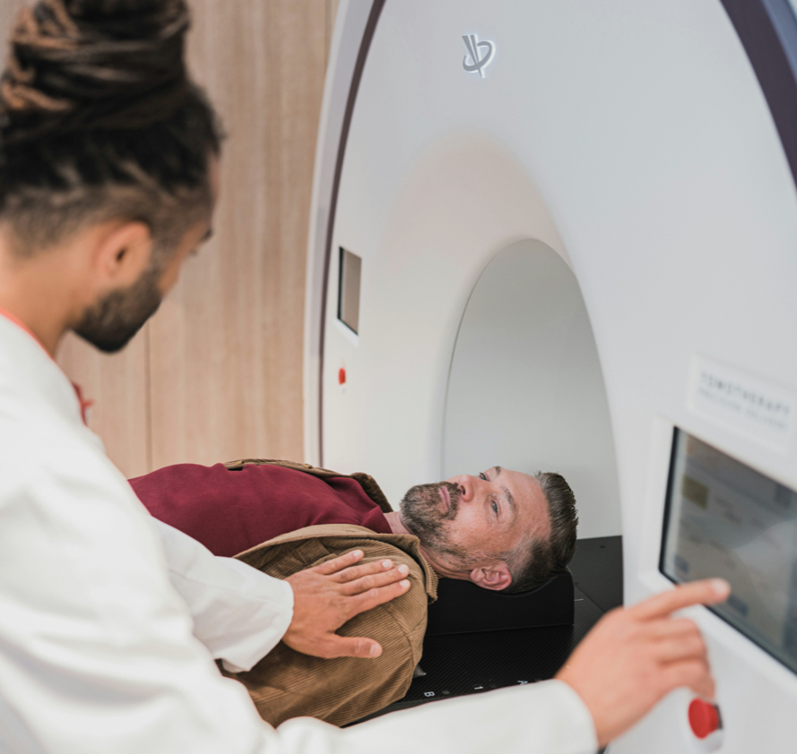
- You'll lie on your back on a couch that slides into the MRI machine.
- A contrast dye may be injected into your arm to enhance images of the prostate.
- Possible mild side effects from the dye: nausea, headache, warmth, metallic taste, dizziness.
- Stay as still as possible (scan takes about 10 minutes).
- The radiographer will monitor you throughout, and you can communicate via an intercom.
- You may be asked to hold your breath at times.
- The MRI machine makes a loud clanging sound—headphones will be provided for hearing protection.
Getting the results
After your mpMRI, a specialist prostate radiologist assigns a PI-RADS or Likert score from 1 to 5: 1–2 means cancer needing treatment is unlikely; 3 is uncertain; 4–5 means it is likely to very likely. At The Focal Therapy Clinic your scan is reported by a consultant radiologist to recognised quality standards (PI-QUAL).
A radiologist will review your MRI images and assign a score from 1 to 5, known as your PI-RADS or Likert score. This score indicates the likelihood of cancer in your prostate, with 1 being low risk and 5 being high risk.
| PI-RADS or Likert score | What it means |
|---|---|
| Score 1 | It's very unlikely that you have prostate cancer that needs to be treated |
| Score 2 | It's unlikely that you have prostate cancer that needs to be treated |
| Score 3 | It isn't possible to tell from the scan whether you have prostate cancer that needs to be treated - you may hear this called a borderline result. |
| Score 4 | It's likely that you have prostate cancer that needs to be treated |
| Score 5 | It's very likely that you have prostate cancer that needs to be treated. |
The PI-QUAL scoring system used to assess prostate MRI reporting quality was developed by Dr Francesco Giganti (first author), with Dr Clare Allen and Dr Aqua Asif of The Focal Therapy Clinic among its co-authors (Radiology, 2023).

Fusion MRI
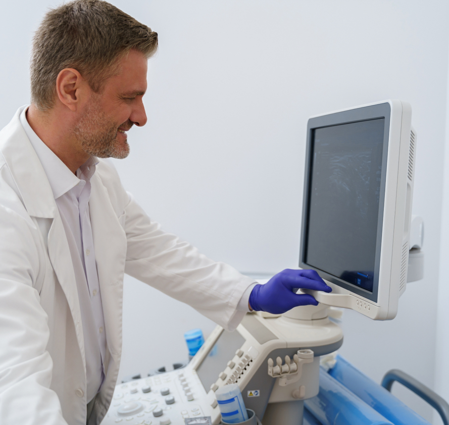
An MRI-ultrasound fusion biopsy overlays your mpMRI onto live ultrasound so the needle targets the exact suspicious area, rather than sampling the prostate at random. This targeted approach — established by the PRECISION and VISION trials — finds more significant cancers and misses fewer, while sparing men with low-risk findings an unnecessary biopsy.
Fusion MRI is an advanced technique that combines MRI scans with live ultrasound to guide prostate cancer treatments.
In a fusion MRI, our expert radiographer marks your prostate, your tumour and lesions, your seminal vesicles and your urethra on your scan results. In a fusion biopsy, a grid is also added. During your biopsy, your MRI scan is overlayed onto your live ultrasound, and a grid-shaped needle guide is used to accurately sample the target area. With a transperineal fusion MRI-guided biopsy, there is very little downtime or soreness afterwards, and it’s much more accurate in confirming a suspected diagnosis of prostate cancer.
During fusion focal therapy, the MRI scan is overlayed onto your live ultrasound and this is used to guide your treatment; for HIFU, this is an ultrasound probe, and for NanoKnife, it is needles. This allows your surgeon to target your tumour very accurately, increasing the effectiveness of your treatment and reducing the risk of damaging any other tissue.
If your MRI and biopsy confirm you're suitable, focal therapy (HIFU or NanoKnife) can treat the cancer while sparing healthy tissue — see focal therapy treatment options.
- Improved Tumour Detection – More accurate than ultrasound alone
- Greater Targeting Precision – Helps specialists precisely pinpoint tumours
- Reduces Risk of Missing Cancerous Tissue – Ensures comprehensive tumour destruction
How Is an Ultrasound-MRI Fusion Prostate Biopsy Done?
You first receive a prostate MRI. The radiologist reviews the images for areas that are suspicious for prostate cancer.
Next, doctors perform an ultrasound test. Your doctor provides local anaesthesia for your comfort and inserts a small ultrasound probe into the rectum.
The fusion biopsy technology combines your MRI images with the ultrasound image in real time. This fusion (blending) of images creates a clearer picture to pinpoint any suspicious areas in the prostate.
Your doctor uses the ultrasound and prostate MRI images to collect the prostate biopsy (tissue sample) with a needle. Only urologists with special training and knowledge of prostate cancer perform this procedure.
“I didn’t have a multi parametric [MRI]. It was a conventional imaging, and that didn't show anything on the normal MRI…..it was from the biopsy how I was diagnosed.The biopsy showed that I had prostate cancer, and fortunately, because I caught it early, it was Gleason six, which is the early stages.”
Alphonso Archer
The Focal Therapy Clinic patient
Questions to ask your doctor or nurse

Get Expert Advice & The Latest Research
Subscribe to our newsletter to receive the latest updates, expert insights, and breakthrough research on prostate cancer-delivered straight to your inbox.
Frequently asked questions

Learn more about focal therapy for prostate cancer

Survey of Prostate Cancer Patients Reveals Severe Knowledge Gaps Among Men

How Accurate is a Urine test for Prostate Cancer – and can it replace PSA or MRI?

Is Testosterone Replacement Therapy Safe After Prostate Cancer Treatment?
Sources
Medically reviewed by Dr Clare Allen, Consultant Radiologist (mpMRI pioneer; PROMIS lead radiologist).
Evidence: Kasivisvanathan V, … Asif A, … Moore CM. VISION: MRI-targeted vs standard biopsy. European Urology 2024;87(5):512–523 (doi:10.1016/j.eururo.2024.08.022) — co-authored by FTC's Dr Aqua Asif. Giganti F, Ng A, Asif A et al. PI-QUAL. Radiology 2023 (doi:10.1148/radiol.231130) — co-authored by FTC's Dr Aqua Asif and Dr Francesco Giganti. Ahmed HU et al. PROMIS. Lancet 2017. Kasivisvanathan V et al. PRECISION. NEJM 2018.
Guideline: NICE NG131.
Subscribe to our newsletter
Get expert advice & the latest prostate cancer research - straight to your inbox.