
At a Glance
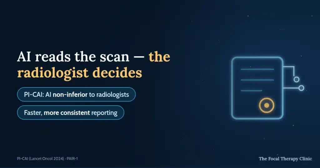
Artificial intelligence is moving from concept to practical tool in prostate cancer diagnosis. AI software now supports radiologists reading prostate MRI — automatically outlining the gland, highlighting suspicious areas and helping apply PI-RADS scores consistently — making reporting faster and more reproducible between hospitals.¹ ⁴ Crucially, current tools assist rather than replace the radiologist, whose clinical judgement remains irreplaceable. For patients, the payoff is quicker, more consistent diagnosis — and sharper targeting for treatments like focal therapy.
Key takeaways:
- Why AI is needed — after PROMIS and PRECISION put MRI before biopsy, scan volumes surged and radiology workloads strained; AI addresses exactly that pressure.¹ ²
- What it does — gland segmentation (volume and PSA density), lesion detection, and PI-RADS scoring support.
- Validated performance — the international PI-CAI study found AI non-inferior to radiologists at detecting significant cancer on MRI.⁴
- Radiologists stay in charge — AI is a second reader; automation bias is a known risk that human oversight controls.
- It feeds focal therapy — precise lesion mapping supports candidate selection and targeting for HIFU and NanoKnife.
The use of MRI in the prostate cancer pathway grew dramatically after PROMIS and PRECISION showed that MRI before biopsy finds more significant cancers and spares many men invasive procedures.¹ ² Guidelines now recommend upfront MRI — which surged scan numbers and strained radiology departments with workload and reader variability. AI offers solutions to exactly these challenges. Almar van Loon, Director of Customer Success at Quantib — developer of the Quantib Prostate tool — works with clinicians on precisely this problem: supporting radiologists reading complex prostate MRI at scale.
What is AI in MRI?
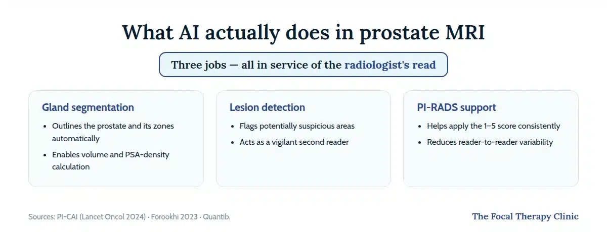
AI in prostate MRI means software built on machine-learning and deep-learning algorithms trained on thousands of prostate MRI scans with known outcomes, learning to recognise the patterns associated with cancer. Key functions:
- Gland segmentation — automatically outlining the prostate and its zones; essential for calculating volume and PSA density (PSAD), a key risk indicator alongside PSA.
- Lesion detection — identifying potentially suspicious areas, sometimes highlighting them visually.
- PI-RADS support — helping radiologists apply the 1-to-5 Prostate Imaging Reporting and Data System score consistently, automating measurements and structuring reports.
Current tools are designed to complement, not replace, the radiologist — automating time-consuming tasks and standardising PI-RADS application, which otherwise varies between readers. The result: more reproducible reports, and more radiologist time for complex interpretation.
How AI improves prostate MRI
A short explainer of precision imaging in prostate cancer — how modern scanning pinpoints tumours so treatment can target only the cancer.
Automating manual tasks like volume calculation saves real time — radiologists using tools like Quantib Prostate report faster reporting and more cases handled per day. Faster reporting means quicker results, shorter waits for biopsy or treatment planning, and relief for busy systems like the NHS.
Structured, visual AI-assisted reports also improve communication between radiology and urology, making MDT meetings more efficient — and standardised workflows reduce the variability between readers and hospitals that has dogged prostate MRI.⁶
Key benefits of AI in scans
- Workflow efficiency — segmentation and calculation automated; backlogs reduced.
- Enhanced communication — standardised visual reports clarify findings for the whole clinical team and support targeted-biopsy planning.
- Diagnostic confidence — AI as a “second reader” can improve accuracy and support less experienced readers, especially on challenging scans.⁶
Limitations and concerns
Adoption faces real challenges. Automation bias — over-relying on AI output without critical appraisal — is a known risk; radiologists must evaluate AI suggestions in full clinical context. Validation across diverse populations, scanner types and hospitals is paramount — studies like PAIR-1, testing AI on real-world NHS data, exist precisely for this.³ Integration with hospital systems (PACS) raises data-governance, privacy and training questions; the “black box” nature of some models can hinder trust; and regulatory approval (CE mark, FDA clearance) plus cost-effectiveness remain hurdles to wide adoption.
AI use in clinics today
AI is steadily moving from research into practice. Commercial tools such as Quantib Prostate are used in hospitals across Europe and the US, with studies showing improved detection sensitivity and support for less experienced readers.⁶ In the UK, NHS trusts are actively evaluating AI: the PAIR-1 study validated Lucida Medical’s Pi software across multiple diverse NHS sites,³ and trusts including Somerset and Leeds are piloting tools to speed the diagnostic pathway. The landmark international PI-CAI study — nearly 10,000 scans — found AI non-inferior to radiologists for detecting clinically significant cancer on MRI.⁴
Get Expert Advice & The Latest Research
Subscribe to our newsletter to receive the latest updates, expert insights, and breakthrough research on prostate cancer-delivered straight to your inbox.
Why radiologists still matter
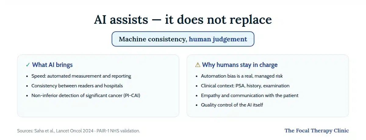
AI assists; it does not replace. As Almar van Loon emphasised, the goal is synergy between human expertise and machine intelligence. The radiologist synthesises MRI findings with clinical history, PSA, examination and other data, and interprets ambiguous patterns in context — which AI currently cannot. Healthcare also needs human connection: empathy, nuance and reassurance when discussing findings with patients. Radiologists oversee AI quality control too. By automating the routine, AI arguably makes radiologist expertise more vital, not less.
The future of AI in imaging
Current applications are just the start. Ahead:
- Early detection and screening — faster, more consistent scan analysis could make MRI-based screening programmes feasible. As Dr Christos Mikropoulos notes, “Early diagnosis is key in prostate cancer”.
- Risk stratification — radiomics combined with clinical and genomic data may distinguish indolent from significant disease more accurately.
- Predicting treatment response — AI may help forecast response to radiotherapy or hormone therapy, personalising choices.
- Monitoring — tracking subtle tumour changes for men on active surveillance or after treatment.
AI’s precise lesion segmentation directly supports targeted treatment planning: detailed tumour location and boundary information is exactly what selecting and targeting focal therapy — HIFU (NICE HTG667) or NanoKnife (NICE HTG688) — requires. The future likely blends imaging, pathology, genetics and clinical history into personalised risk assessment and management for each patient.
AI is one thread of a bigger story — see how the precision-imaging revolution is changing prostate cancer diagnosis.
Frequently Asked Questions
Will AI read my prostate MRI instead of a doctor?
No — current tools assist the radiologist, who reviews everything the AI flags within your full clinical picture. Regulation and good practice keep a human expert responsible for your report.
Does AI actually perform as well as radiologists?
In the international PI-CAI study of nearly 10,000 scans, AI was non-inferior to radiologists at detecting clinically significant cancer on MRI⁴ — as a second reader alongside a human, it can raise overall consistency.
Is AI being used in the NHS now?
It is being actively evaluated: the PAIR-1 study validated one tool across diverse NHS sites, and several trusts are piloting AI to speed their diagnostic pathways.³
How does AI help with focal therapy?
By mapping lesion location, size and boundaries precisely, AI-assisted MRI supports both the decision about whether focal therapy is suitable and the accurate targeting of treatment — treating the cancer while sparing healthy tissue.
This content is not intended to be a substitute for professional medical advice, diagnosis, or treatment. Always seek the advice of your urologist or other qualified health provider with any questions you may have regarding a medical condition.
References
- Ahmed HU, et al. (PROMIS Study Group). Diagnostic accuracy of multi-parametric MRI and TRUS biopsy in prostate cancer (PROMIS). Lancet. 2017;389(10071):815–822.
- Kasivisvanathan V, et al. (PRECISION Study Group). MRI-Targeted or Standard Biopsy for Prostate-Cancer Diagnosis. N Engl J Med. 2018;378(19):1767–1777.
- Giganti F, et al. AI-powered prostate cancer detection: a multi-centre, multi-scanner validation study (PAIR-1). ECR 2025; Lucida Medical PAIR-1 summary. https://lucidamedical.com/pi-expert-performance-with-nhs-patient-data/
- Saha A, et al. (PI-CAI Consortium). Artificial intelligence and radiologists in prostate cancer detection on MRI (PI-CAI): an international, paired, non-inferiority, confirmatory study. Lancet Oncol. 2024;25(7):879–887.
- NHS AI Lab — programme archive: NHS England Digital, AI knowledge repository. https://digital.nhs.uk/services/ai-knowledge-repository
- Forookhi A, et al. Bridging the experience gap in prostate multiparametric MRI using artificial intelligence. Eur J Radiol. 2023;161:110749.
- Quantib. Quantib Prostate product overview; RadNet FDA clearance announcement (2023). https://www.quantib.com/en/solutions/quantib-prostate