
At a Glance
Multiparametric MRI (mpMRI) is now the standard first-line imaging tool for prostate cancer diagnosis in the UK, recommended by NICE guideline NG131. A high-quality prostate MRI helps your clinical team detect clinically significant cancers, avoid unnecessary biopsies, and plan targeted treatments such as focal therapy — preserving quality of life.
- Better cancer detection — mpMRI identifies clinically significant cancers while reducing overdiagnosis of low-risk disease
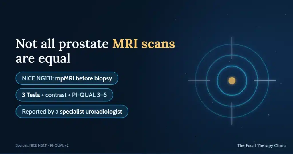
- Not all scans are equal — magnet strength, contrast protocol, PI-QUAL score and who reports the scan all determine whether your imaging is good enough to plan treatment
- Guides treatment planning — accurate imaging determines eligibility for focal therapies such as HIFU (NICE HTG667) and NanoKnife IRE (NICE HTG688)
- Preserves quality of life — MRI-guided focal therapy at The Focal Therapy Clinic achieves 90%+ sexual function preservation and 97% urinary continence (FTC audit, n=265)
- Expert interpretation matters — scans should be read by specialist uroradiologists for the most accurate results
Why Is Prostate MRI Now the Standard of Care?
Multiparametric MRI (mpMRI) has transformed prostate cancer diagnosis and is now recommended by NICE (guideline NG131) as the first-line imaging tool before biopsy. A high-quality prostate MRI allows clinicians to detect clinically significant cancers, avoid unnecessary biopsies, and plan targeted treatments — including focal therapy — with far greater precision than the traditional PSA-only pathway.
Professor Francesco Giganti, Professor of Radiology at University College London and Consultant Radiologist (GMC: 7471579), is a leading authority on prostate imaging with over 200 peer-reviewed publications. In a recent OnFocus podcast, he discussed how mpMRI has become essential to the prostate cancer diagnostic pathway.
How Has MRI Transformed Prostate Cancer Diagnosis?
Multiparametric MRI (mpMRI) has replaced the traditional PSA-then-biopsy approach as the recommended first step in prostate cancer diagnosis. NICE guideline NG131 now recommends that all men with suspected prostate cancer receive an mpMRI before any biopsy, allowing clinicians to identify clinically significant cancers while reducing unnecessary biopsies and the overdiagnosis of low-risk disease.
Before mpMRI became standard, diagnosis relied heavily on PSA blood tests followed by systematic biopsies — an approach that sometimes missed significant cancers or detected clinically insignificant ones, leading to overtreatment. As Professor Francesco Giganti explains, mpMRI provides detailed images of the prostate that allow clinicians to assess the size, location, and characteristics of suspicious lesions before any tissue sampling takes place.
| Aspect | Traditional PSA-biopsy pathway | mpMRI-first pathway (NICE NG131) |
|---|---|---|
| First step | PSA blood test → systematic biopsy | PSA blood test → mpMRI before biopsy |
| Biopsy targeting | Systematic (random sampling) | MRI-targeted (guided to suspicious areas) |
| Overdiagnosis risk | Higher (detects low-risk cancers) | Lower (focuses on clinically significant disease) |
| Missed cancers | More likely (anterior tumours often missed) | Less likely (MRI visualises the entire gland) |
| NICE recommendation | No longer recommended as first line | Recommended standard (NG131) |
Source: NICE guideline NG131 — Prostate cancer: diagnosis and management.
How Does MRI Guide Prostate Cancer Treatment Decisions?
Accurate mpMRI imaging is essential for determining which men are suitable for focal therapy — a targeted approach that treats only the cancerous area of the prostate while preserving surrounding healthy tissue. At The Focal Therapy Clinic, every patient undergoes detailed MRI review as part of a multidisciplinary assessment before any treatment recommendation is made.
Professor Francesco Giganti emphasises that the quality of the MRI directly influences treatment planning. A high-resolution scan allows the clinical team to map the precise location, size, and extent of the tumour — information critical for planning targeted treatments such as HIFU (High-Intensity Focused Ultrasound), approved by NICE under HTG667, or NanoKnife IRE, approved under NICE HTG688.
In our clinic’s experience of 2,500+ focal therapy procedures, MRI-guided treatment planning contributes to outcomes including 90%+ sexual function preservation and 97% urinary continence (FTC audit, n=265).
What Are the Benefits of MRI-Guided Focal Therapy?
Mr Marc Laniado explains MRI-ultrasound fusion technology and how precision imaging underpins focal therapy’s success rates.
MRI-guided focal therapy treats only the cancerous area of the prostate, resulting in fewer side effects and faster recovery than whole-gland treatments such as radical prostatectomy or radiotherapy. At The Focal Therapy Clinic, our patient data demonstrates the benefits of precise MRI-guided treatment planning.
| Outcome | Focal therapy (FTC data) | Radical prostatectomy |
|---|---|---|
| Sexual function preserved | 90%+ | 30–70% |
| Urinary continence | 97% (FTC audit, n=265) | 80–95% |
| Recovery time | 1–2 weeks | 4–6 weeks |
| Hospital stay | Day-case (home same day) | 1–3 nights |
| NICE status | HIFU: HTG667 · NanoKnife: HTG688 | Standard care |
Source: FTC one-year outcome audit (n=265) and published literature.
As Professor Francesco Giganti notes, high-quality MRI is the foundation of these outcomes — precise imaging allows the clinical team to target treatment accurately, preserving healthy tissue and maintaining quality of life.
“The quality of the MRI directly determines the quality of the treatment plan. A high-resolution multiparametric scan allows us to map the tumour precisely, which is essential for focal therapy to achieve its potential.” — Professor Francesco Giganti, Professor of Radiology, University College London (GMC: 7471579)
Why Prostate MRI Quality Varies — and Why It Matters
Mr Marc Laniado, Consultant Urological Surgeon and Prostate Cancer Lead at Wexham Park Hospital, identifies a fundamental problem in UK prostate cancer care: “one man’s prostate MRI is not another man’s prostate MRI.” While mpMRI is now standard practice under NICE NG131, dramatic quality variations affect which treatments patients can access.
“We see a huge variation in the scanners, the administration of contrast and the interpretation of the scans, and that poses major and significant challenges to what can be offered to patients.” — Mr Marc Laniado, Consultant Urological Surgeon
Poor imaging can send patients down the wrong path in both directions: advised towards radical surgery when focal therapy would suffice, or reassured onto active surveillance when significant disease has been missed. Poor initial imaging can also eliminate future focal therapy options.
The precision difference in practice. Consider a typical 60-year-old with Gleason 3+4=7 (Grade Group 2) cancer and PSA 6 — “intermediate risk” on paper. That single label can hide two very different situations:
- Patient A: small, focal cancer, no urinary symptoms → suitable for focal ablation preserving function
- Patient B: large cancer occupying most of the prostate, with urinary symptoms → needs whole-gland treatment such as radical prostatectomy
“Precision diagnostics is really about understanding the severity of the cancer, but also knowing much more about its location, the volume, the grade, its position than was typically given in the past.” — Mr Marc Laniado
Only high-quality imaging can tell these two men apart — and give each the treatment he actually needs.
Technical Standards That Matter
Understanding the specifications empowers you to advocate for diagnostic-quality imaging:
| Parameter | Optimal standard | Suboptimal | Clinical impact |
|---|---|---|---|
| Magnet strength | 3 Tesla | ≤1.5 Tesla | Higher resolution enables focal therapy planning |
| Contrast protocol | mpMRI with gadolinium contrast | Biparametric (no contrast) | Essential for targeting and post-treatment monitoring |
| PI-QUAL score | 3 (diagnostic quality) | 1–2 (poor quality) | Determines whether findings can be trusted |
| Reporting | Dedicated uroradiologist | General radiologist | Affects interpretation reliability |
Why contrast matters. Contrast agents reveal blood-flow patterns that help distinguish cancerous from healthy tissue — “cancers often take up more blood flow than non-cancers”. Contrast is essential for precise focal therapy targeting, for distinguishing cancer from benign enlargement, and — critically — for the follow-up scans that show how effective focal treatment has been. Without contrast you have a biparametric, not multiparametric, MRI.
PI-QUAL scoring. The PI-QUAL v2 system grades scan quality from 1 (poor) to 3 (optimal); scores of 2 or above are considered diagnostic quality. The standard is not yet consistently applied across UK hospitals — which is exactly why it is worth asking about.
The Modern Diagnostic Pathway
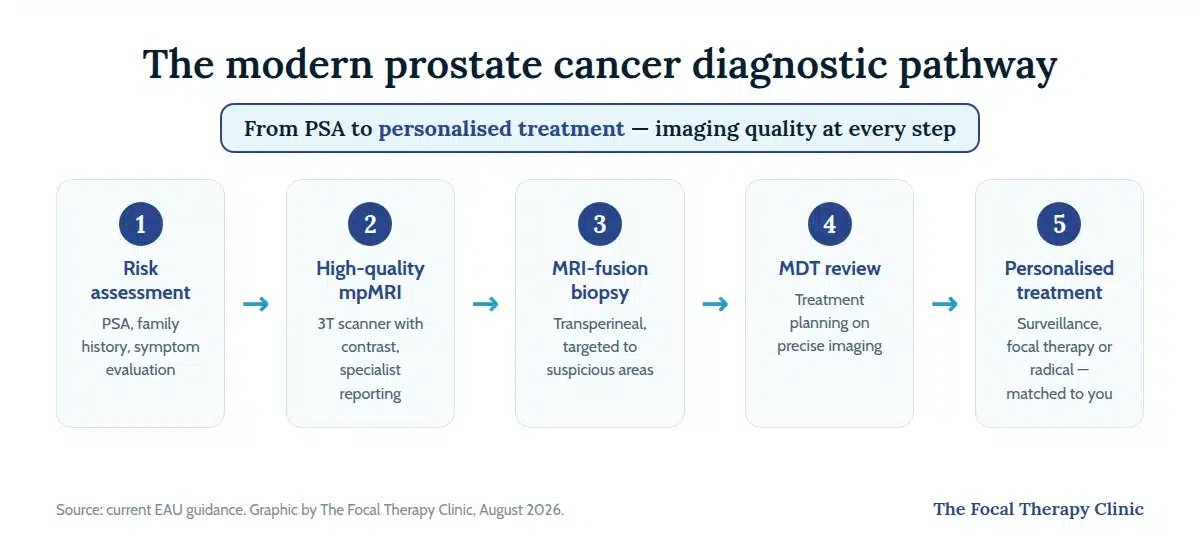
Best-practice sequence, in line with current EAU guidance:
- Risk assessment — PSA, family history, symptom evaluation
- High-quality mpMRI — 3T scanner with contrast, specialist reporting
- MRI-fusion targeted biopsy — transperineal approach, guided to suspicious areas
- Multidisciplinary review — treatment planning based on precise imaging
- Personalised treatment — from active surveillance to focal therapy to radical intervention, matched to the individual
To see where each of these steps sits in the journey from first test to treatment decision, read our full prostate cancer guide.
Get Expert Advice & The Latest Research
Subscribe to our newsletter to receive the latest updates, expert insights, and breakthrough research on prostate cancer-delivered straight to your inbox.
What Should You Ask About MRI Quality at Your Hospital?
Not all prostate MRI scans are equal. Ask specific questions about the equipment and reporting at your treatment centre, because scan quality directly affects diagnostic accuracy and treatment planning:
- Is it multiparametric? — A standard MRI is not sufficient. You need mpMRI with T2-weighted, diffusion-weighted and contrast-enhanced sequences
- What magnet strength? — 3-Tesla scanners provide higher resolution than 1.5-Tesla for prostate imaging
- Who reports the scan? — It should be a specialist uroradiologist experienced in prostate MRI, not a general radiologist
- Is PI-RADS scoring used? — PI-RADS standardises how findings are reported, scoring lesions from 1 (very low risk) to 5 (very high risk)
- What PI-QUAL score does your radiology unit achieve on audit? — 1 – 3 is diagnostic quality
- Will my biopsy use MRI-ultrasound fusion for targeting accuracy?
- Can I get a second opinion? — If you are unsure about your results, you have the right to seek one from a specialist centre
At The Focal Therapy Clinic, all MRI scans are reviewed by specialist uroradiologists including Dr Clare Allen (FRCR, GMC: 3108389), ensuring accurate PI-RADS scoring and detailed tumour mapping before any treatment decision is made.
Frequently Asked Questions
Do I really need contrast for a prostate MRI?
Yes. Contrast highlights blood-rich tumours and is essential for assessing treatment success after focal therapy. Without contrast you receive a biparametric rather than multiparametric MRI.
What PI-QUAL score should my scan achieve?
Aim for 2 -3 ; below 2 the image quality is too poor for confident diagnosis and treatment planning.
Can mpMRI replace a biopsy entirely?
Not yet. A negative high-quality mpMRI lowers the probability of significant cancer, but guidelines still recommend targeted biopsy where clinical suspicion remains.
How does scan quality affect my treatment options?
Poor imaging may point towards radical treatment when focal therapy would suffice — or miss significant disease. Quality imaging is what makes personalised treatment selection possible.
What if my local hospital doesn’t meet these standards?
Consider referral to a specialist centre for repeat imaging, particularly if you are considering focal therapy. You are entitled to a copy of your images for a second opinion.
What Should You Do Next?
If you have been diagnosed with prostate cancer or are awaiting further investigation, ensuring access to a high-quality multiparametric MRI is one of the most important steps you can take. At The Focal Therapy Clinic, your MRI is reviewed by specialist uroradiologists as part of a comprehensive assessment to determine whether focal therapy may be suitable for you.
- Request your MRI images — you are entitled to a copy of your scan for a second opinion
- Ask about mpMRI — ensure your scan is multiparametric, not a standard MRI
- Book a consultation — our team can review your existing MRI and advise on next steps, including whether focal therapy is an option
About the Clinicians in This Article
Professor Francesco Giganti — Professor of Radiology and Consultant Radiologist, University College London / UCLH. Expertise: prostate MRI, active surveillance imaging, MRI-targeted biopsies. Over 200 peer-reviewed papers on prostate imaging.
Mr Marc Laniado — Consultant Urological Surgeon at The Focal Therapy Clinic and Prostate Cancer Lead at Wexham Park Hospital, Frimley Health. Qualifying in 1989 and a consultant since 2002, he has pioneered imaging-led diagnostics and championed patients’ rights to informed treatment choices. His expertise spans advanced MRI interpretation, fusion biopsy techniques and focal therapy delivery.
Listen to the Full Podcast
Hear Professor Francesco Giganti discuss the role of MRI in prostate cancer diagnosis and treatment on the OnFocus podcast:
References
- NICE guideline NG131 — Prostate cancer: diagnosis and management.
- NICE HealthTech guidance HTG667 — Focal therapy using high-intensity focused ultrasound for localised prostate cancer.
- NICE HealthTech guidance HTG688 — Irreversible electroporation for treating prostate cancer.
- PI-QUAL v2 — prostate MRI quality assessment.
- EAU guidelines on prostate cancer.
- The Focal Therapy Clinic one-year outcome audit (n=265).
This content is not intended to replace professional medical advice. Always consult your clinical team about your individual circumstances.