
At a Glance
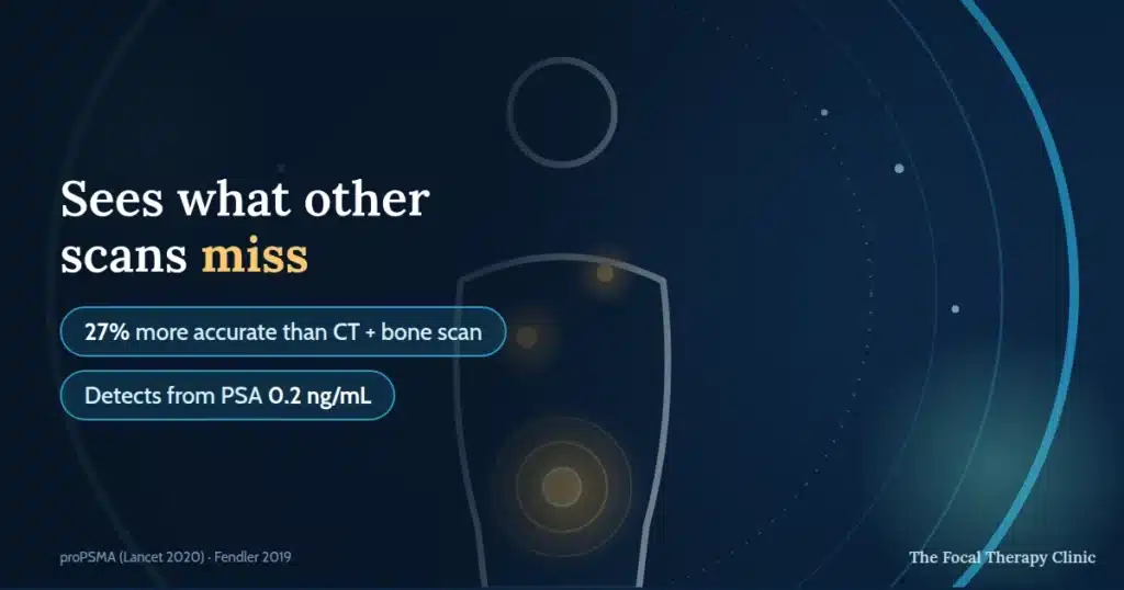
A PSMA PET scan is a specialised imaging test that detects prostate cancer cells throughout the body by targeting prostate-specific membrane antigen (PSMA), a protein overexpressed on prostate cancer cells. In the landmark proPSMA trial it was 27% more accurate than CT and bone scan combined, and changed management in 28% of cases. It can detect disease at PSA levels as low as 0.2 ng/mL. At The Focal Therapy Clinic, PSMA PET scanning is part of our diagnostic workup to confirm whether cancer is truly localised before recommending focal therapy.
Key takeaways
- Targets cancer cells directly — uses a radioactive tracer that binds to PSMA, a protein overexpressed on prostate cancer cells.
- Detects spread earlier — identifies metastases and recurrence at PSA levels as low as 0.2 ng/mL, when other scans may miss them.
- Changes treatment plans — the landmark proPSMA trial showed management changed in 28% of cases.
- Guides focal therapy decisions — helps confirm that cancer is localised, supporting treatments like HIFU (NICE HTG667) or NanoKnife IRE (NICE HTG688).
- Safe and well-tolerated — an outpatient procedure taking 2–3 hours, with the tracer cleared naturally through urine within 24 hours.
What is a PSMA PET scan?
A PSMA PET scan is a whole-body imaging test that uses a radioactive tracer to find prostate cancer cells by binding to PSMA, a protein found in high concentrations on the surface of prostate cancer cells.
A standard PET scan often uses FDG, a general tracer that highlights areas of high metabolic activity, while a PSMA PET scan uses a prostate-specific tracer. This makes it more useful for prostate cancer staging, biochemical recurrence, and treatment planning. It can target cancer cells directly, detect spread earlier, change treatment plans in 28% of cases, guide focal therapy decisions, and is usually safe and well tolerated.
Major European guidelines now recognise PSMA PET imaging as a leading option for prostate cancer imaging, especially in higher-risk cases. EAU-EANM-ESTRO-ESUR-ISUP-SIOG Guidelines 2024 support its use where accurate staging may change treatment decisions.
What PSMA is and why it matters
PSMA, or prostate-specific membrane antigen, is a protein found on the surface of all prostate cells but is produced in dramatically higher quantities by prostate cancer cells — published reviews cite up to 100 to 1,000 times more PSMA than healthy tissue. This overexpression gives doctors a biological signal that radioactive tracers can lock onto. The scan then highlights areas where prostate cancer cells may be active.
PSMA levels also tend to rise as prostate cancer becomes more aggressive, according to Wright et al., Urologic Oncology 1995. This clinical insight is what makes PSMA PET imaging more accurate than conventional scans for many men. A PSMA PET scan prostate cancer assessment can therefore help doctors see whether cancer is still localised or has spread.
Why is PSMA important for finding prostate cancer?
Because the PSMA PET tracer binds specifically to prostate cancer cells, it can identify small deposits of disease that CT scans, bone scans, and standard PET scans would miss — particularly when PSA levels are low. Published research shows PSMA PET can detect cancer recurrence at PSA levels as low as 0.2 ng/mL. This is especially valuable for men with a rising PSA after previous treatment. At The Focal Therapy Clinic, early and accurate detection supports more treatment options — including focal therapies that preserve quality of life.
How does a PSMA PET scan work?
Mr Marc Laniado explains MRI-ultrasound fusion technology and how precision imaging underpins focal therapy’s success rates.
A PSMA PET scan is a straightforward outpatient procedure taking approximately two to three hours from arrival to completion, involving a small injection of a radioactive tracer followed by a whole-body PET scan. The tracer circulates for about 60 minutes, binds to PSMA-expressing cells, and the scan itself usually takes 20 to 30 minutes. The images are then reviewed by specialists to identify where prostate cancer cells may be present.
What to expect on the day
On the day of your scan, you will receive a small injection of the PSMA tracer, typically Gallium-68 PSMA-11 or Fluorine-18 DCFPyL, into a vein in your arm, then rest for approximately 60 minutes while the tracer circulates. You will then lie on a table that slides through the PET scanner for around 20 to 30 minutes. The scan is painless and well tolerated by most patients. Patients often ask how long does a PSMA PET scan take, and the full appointment is usually two to three hours.
Preparation tips:
- Wear metal-free clothing, such as jogging trousers and a plain T-shirt
- Avoid jewellery, belts, watches, glasses, and other metal items
- Tell staff if you feel anxious or claustrophobic, as medication can be provided
The tracer clears from the body through urine within 24 hours. The radiation dose is comparable to other routine imaging tests.
How the tracer finds cancer
The PSMA tracer consists of a molecule that binds specifically to PSMA proteins on prostate cancer cells, paired with a small amount of radioactive material that the PET scanner detects. After injection, the tracer enters the bloodstream and attaches to PSMA-expressing cells anywhere in the body. The scanner detects this signal and creates a 3D map showing where prostate cancer cells may be located — including deposits too small for CT, MRI, or bone scan to identify.
Gallium-68 PSMA-11 and Fluorine-18 DCFPyL are two commonly used tracers. The EANM/SNMMI Procedure Guideline 2017 by Fendler et al. supports standardised use of PSMA PET imaging in prostate cancer assessment.
When is a PSMA PET scan recommended?
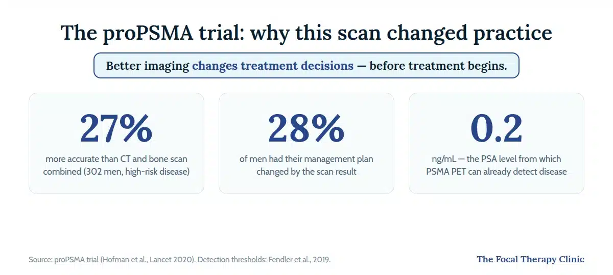
A PSMA PET scan is most commonly recommended in three clinical scenarios: initial staging of intermediate or high-risk newly diagnosed prostate cancer, detecting biochemical recurrence when PSA rises after treatment, and planning treatment, including assessing candidacy for focal therapy.
- Initial staging of newly diagnosed prostate cancer: PSMA PET is most informative for men with higher-risk features, including Gleason score 7 or above, PSA above 10 ng/mL, or clinical stage T2c or above. NICE NG131 sets out how prostate cancer should be diagnosed and managed. A PSMA PET scan may help confirm whether cancer is confined to the prostate or has spread.
- Detecting recurrence after treatment: A rising PSA after surgery or radiotherapy is one of the strongest reasons to consider PSMA PET imaging. The scan can help identify whether cancer has returned locally or spread to distant sites, with useful detection rates even at low PSA levels.
- Treatment planning, including focal therapy candidacy: Confirming that cancer is genuinely localised is essential before recommending focal therapy. This matters because treatments that target only the cancerous area should not be used when cancer has already spread outside the prostate. PSMA PET results can therefore help guide whether local, focal, or wider treatment is most appropriate.
Not every man with prostate cancer requires a PSMA PET scan — a consultant will advise based on individual circumstances.
How accurate is a PSMA PET scan?
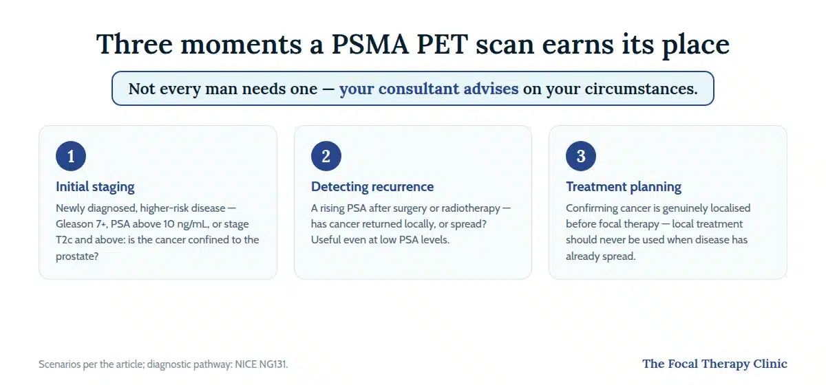
PSMA PET scans are significantly more accurate than conventional bone scans and CT for detecting prostate cancer spread: the landmark proPSMA trial (302 men with high-risk prostate cancer) demonstrated 27% greater accuracy than standard imaging and changed management decisions in 28% of cases. Conventional imaging becomes unreliable at low PSA levels, which is exactly where PSMA PET keeps its sensitivity — this is why better imaging directly affects treatment selection. Men often ask how accurate is PSMA PET scan because the result may decide whether they are suitable for localised treatment, especially men being assessed for HIFU treatment.
Detection rates by PSA level
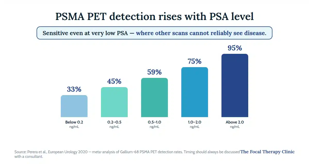
PSMA PET detection rates rise steadily with PSA level, but the scan remains sensitive even at low PSA values where other imaging tests cannot reliably detect disease.
| PSA level (ng/mL) | Detection rate |
|---|---|
| Below 0.2 | 33% |
| 0.2–0.5 | 45% |
| 0.5–1.0 | 59% |
| 1.0–2.0 | 75% |
| Above 2.0 | 95% |
These detection rates come from the Perera et al. meta-analysis in European Urology 2020, a systematic review of Gallium-68 PSMA PET across advanced prostate cancer studies. Timing should always be discussed with a consultant because earlier is not always better. Detection depends on PSA trend, cancer grade, and previous treatment history.
Limitations and false results
PSMA PET scans are highly accurate but not infallible, and clinicians always interpret results in the context of PSA trends, biopsy findings, and other imaging. Some low-grade or neuroendocrine prostate cancers express little PSMA and may be missed. False positives can also occur because PSMA uptake may appear in ganglia, inflammation, or certain non-prostate tissues. These limits are one reason every result needs specialist review before decisions such as NanoKnife IRE treatment are made.
Possible side effects are uncommon and generally mild:
- Injection site discomfort
- Mild allergic reaction
- Rare temporary nausea or tiredness
Get Expert Advice & The Latest Research
Subscribe to our newsletter to receive the latest updates, expert insights, and breakthrough research on prostate cancer-delivered straight to your inbox.
How much does a PSMA PET scan cost in the UK?
A private PSMA PET-CT scan in the UK typically costs around £1,800–£2,500 — an indicative market range, not a Focal Therapy Clinic price — and is free on the NHS for men who meet NHS England’s eligibility criteria.
PSMA PET scans are increasingly available across the UK. Since February 2025, NHS England has routinely commissioned PSMA PET-CT for men with high-risk primary prostate cancer or biochemical recurrence who meet the policy’s eligibility criteria, while private clinics offer self-pay or insurance-covered access at approximately £1,800 to £2,500. NHS waiting times and scanner capacity still vary by region, and men outside the commissioned criteria may not qualify. Private access may be faster and may offer broader eligibility for men needing timely treatment decisions.
Many private health insurance plans now cover PSMA PET scans for high-risk or recurrent disease. The Focal Therapy Clinic offers PSMA PET scans as part of its advanced prostate cancer diagnostic pathway. Men comparing PSMA PET scan UK options should speak to the clinic’s patient coordinator, or read our guide to private prostate cancer diagnostics and what they cost.
Is a PSMA PET scan right for you?
A PSMA PET scan may be appropriate if you have been newly diagnosed with intermediate- or high-risk prostate cancer, if your PSA is rising after previous treatment, or if your clinical team needs to determine whether cancer remains localised before recommending focal therapy. Not every patient requires one — your consultant will advise based on your individual circumstances. If the scan confirms the cancer is confined to the prostate, check whether you are suitable for focal therapy.
“Not every patient is suitable for focal therapy, and we’re transparent about that. We assess every referral carefully with mpMRI and targeted biopsy — and advanced imaging like PSMA PET — before recommending treatment.” — Mr Raj Nigam, Consultant Urological Surgeon (FRCS (Urol), GMC: 3265226)
At The Focal Therapy Clinic, PSMA PET scans are part of our comprehensive diagnostic assessment. Every case is reviewed by our multidisciplinary team — including consultant urological surgeons with collective experience of more than 2,500 focal therapy procedures across six UK cities — to determine the most appropriate treatment pathway.
Questions to raise with your doctor:
- Will the scan change my treatment? — ask whether results would alter the recommended approach.
- Is the timing right? — PSMA PET is most informative when PSA and clinical stage suggest potential spread.
- What happens next? — understand which treatment options become available depending on results.
- Is it covered by insurance? — many private plans now cover PSMA PET for high-risk or recurrent disease.
Frequently Asked Questions
What is a PSMA PET scan for prostate cancer?
A PSMA PET scan is a specialised imaging test that uses a radioactive tracer to detect prostate cancer cells throughout the body. The tracer binds to prostate-specific membrane antigen, or PSMA, a protein overexpressed on prostate cancer cells. This allows clinicians to see where cancer is present.
At what PSA level should a PSMA PET scan be done?
A PSMA PET scan can detect cancer at PSA levels as low as 0.2 ng/mL, though detection rates improve with higher PSA. Most consultants recommend the scan when PSA is rising after treatment or when newly diagnosed cancer is high risk. This includes Gleason 7 or above or PSA above 10 ng/mL.
How accurate is a PSMA PET scan for prostate cancer?
PSMA PET scans are 27% more accurate than conventional bone scans and CT for staging prostate cancer, according to the proPSMA trial by Hofman et al., Lancet 2020. The scan is highly accurate, but it can still miss tumours with low PSMA expression. It can also occasionally flag benign conditions.
What is the difference between a PET scan and a PSMA PET scan?
A standard PET scan uses a general tracer, usually FDG, that highlights areas of high metabolic activity throughout the body, including inflammation. A PSMA PET scan uses a tracer that specifically targets prostate cancer cells through the PSMA protein. This makes it more accurate for prostate cancer detection, staging, and monitoring.
Is a PSMA PET scan available on the NHS?
Yes, PSMA PET scans are available on the NHS: since February 2025 NHS England has routinely commissioned them for high-risk newly diagnosed prostate cancer and biochemical recurrence after treatment, within set eligibility criteria. Waiting times and capacity vary by region. Private clinics may offer faster access and broader eligibility, with some insurance plans covering the scan.
How long does a PSMA PET scan take?
A PSMA PET scan appointment typically takes two to three hours from arrival to completion. This includes the tracer injection, a 60-minute wait while the tracer circulates and binds to cancer cells, and the scan itself. The scanning stage usually lasts around 20 to 30 minutes.
Is a PSMA PET scan safe?
PSMA PET scans are safe and well tolerated, with a radiation dose comparable to other routine imaging tests. The tracer clears naturally through urine within 24 hours. Side effects are uncommon and generally mild, including occasional injection site discomfort or rare temporary nausea.
How much does a PSMA PET scan cost privately in the UK?
A private PSMA PET-CT scan typically costs around £1,800–£2,500 in the UK — an indicative market range rather than a fixed price. The scan is available free on the NHS within set eligibility criteria (see the NHS question in these FAQs), and some private health insurance policies cover it for high-risk or recurrent disease — check your policy.
Speak to a Focal Therapy Specialist
If you have been told you may need a PSMA PET scan or you are considering focal therapy and want to discuss your imaging options, the Focal Therapy Clinic team can help. The clinic’s consultant urological surgeons use PSMA PET scanning as part of a detailed diagnostic workup, with multidisciplinary team review of every case. The team has collective experience of more than 2,500 focal therapy procedures across six UK cities.
Scans are one input to the decision — data-driven tools like Predict Prostate show how the numbers guide treatment choices.
Questions to ask your consultant:
- Will the scan change my treatment?
- Is the timing right?
- What happens next after the result?
- Is the scan covered by insurance?
Book a consultation to discuss imaging, diagnosis, focal therapy suitability, and next steps.
References
References verified via PubMed.
- Wright GL Jr, Haley C, Beckett ML, Schellhammer PF. Expression of prostate-specific membrane antigen in normal, benign, and malignant prostate tissues. Urol Oncol. 1995;1(1):18–28.
- Perera M, Papa N, Roberts M, et al. Gallium-68 prostate-specific membrane antigen positron emission tomography in advanced prostate cancer — updated diagnostic utility, sensitivity, specificity, and distribution of PSMA-avid lesions: a systematic review and meta-analysis. Eur Urol. 2020;77(4):403–417. doi:10.1016/j.eururo.2019.01.049
- Fendler WP, Eiber M, Beheshti M, et al. ⁶⁸Ga-PSMA PET/CT: Joint EANM and SNMMI procedure guideline for prostate cancer imaging. Eur J Nucl Med Mol Imaging. 2017;44(6):1014–1024.
- Hofman MS, Lawrentschuk N, Francis RJ, et al. Prostate-specific membrane antigen PET-CT in patients with high-risk prostate cancer before curative-intent surgery or radiotherapy (proPSMA): a prospective, randomised, multicentre study. Lancet. 2020;395(10231):1208–1216. doi:10.1016/S0140-6736(20)30314-7
- Tilki D, van den Bergh RCN, Briers E, et al. EAU-EANM-ESTRO-ESUR-ISUP-SIOG Guidelines on Prostate Cancer. Part II—2024 Update. Eur Urol. 2024;86(2):164–182. doi:10.1016/j.eururo.2024.04.010
- Fendler WP, Calais J, Eiber M, et al. Assessment of ⁶⁸Ga-PSMA-11 PET accuracy in localizing recurrent prostate cancer. JAMA Oncol. 2019;5(6):856–863.
- Hope TA, Goodman JZ, Allen IE, et al. Meta-analysis of ⁶⁸Ga-PSMA-11 PET accuracy for the detection of prostate cancer validated by histopathology. J Nucl Med. 2019;60(6):786–793. doi:10.2967/jnumed.118.219501
- Rowe SP, Macura KJ, Mena E, et al. PSMA-based PET/CT is superior to conventional imaging for lesion detection in metastatic prostate cancer. Mol Imaging Biol. 2016;18(3):411–419.
- National Institute for Health and Care Excellence. Prostate cancer: diagnosis and management. NICE guideline NG131. London: NICE; 2019 (updated 2021; reviewed 2025). https://www.nice.org.uk/guidance/ng131
- Sengupta S, Asha Krishnan M, Chattopadhyay S, Chelvam V. Comparison of prostate-specific membrane antigen ligands in clinical translation research for diagnosis of prostate cancer. Cancer Rep (Hoboken). 2019;2(4):e1169. (source for the up-to-1,000-fold PSMA overexpression figure)
- NHS England. PSMA radiotracers in PET-CT imaging for individuals with high-risk primary or recurrent prostate cancer: commissioning policy (ref 2307). 27 February 2025. https://www.england.nhs.uk/publication/psma-radiotracers-in-petct-imaging-for-individuals-with-high-risk-primary-or-recurrent-prostate-cancer/
This information is for educational purposes and should not replace professional medical advice. Individual results may vary, and all treatment decisions should be made in consultation with qualified healthcare professionals who can assess your specific circumstances.